Showing posts with label brain mapping. Show all posts

Tuesday, July 10, 2007

Introducing the Brain Maps API


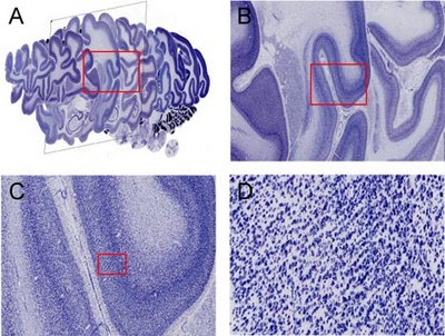
The Brain Maps API lets you embed Brain Maps in your own web pages with JavaScript. Future versions will enable you to add overlays to brain maps (including markers and polylines) and display shadowed "info windows". The Brain Maps API is a free service, available for any web site that is free to consumers.

Try going to this link to see an example, and once there, try clicking and dragging the image, or clicking on the little tree icon in the upper right. Also, the mouse scroll wheel should zoom you in and out.

Try clicking and dragging the image. Also, the mouse scroll wheel should zoom you in and out.

The code for embedding the Brain Maps API into your own website is simple, requiring just a single line of code:

<iframe src="http://brainmaps.org/ajax-viewer.html?path=http://brainmaps.org/HBP2/c.aethiops/AGM1/AGM1-highres/350/&height=78181&width=92160" width="460" height="267" frameborder="0"></iframe>


It's best to actually download the Brain Maps API and upload the required javascript to your own site, since using just the embedding code above relies on cross-site scripting which is disabled in FireFox.

Additional information is available at http://brainmaps.org/index.php?p=brain-maps-api

Saturday, February 24, 2007

Google Earth for the Brain

- Shawn Mikula

Users of BrainMaps.org have often described it as a Google Maps for the Brain, which is interesting because we have taken Google Maps as an inspiration and a guide for what mapping the brain should be like. In line with this, one of the developers at BrainMaps.org, Issac Trotts, has released a veritable Google Earth for the Brain, called StackVis. In a nutshell, StackVis is a 3D viewer of neuroanatomical sections. But it is revolutionary in the sense that it permits rapid interactive viewing of arbitrarily large images.

Conventional microscopy, electron microscopy, and imaging techniques such as MRI and PET commonly generate large stacks of images of the sectioned brain. In other domains, such as neurophysiology, variables such as space or time are also varied along a stack axis. Digital image sizes have been progressively increasing and in virtual microscopy, it is now common to work with individual image sizes that are several hundred megapixels and several gigabytes in size. The interactive visualization of these high-resolution, multiresolution images in 3D has not been possible, until now. StackVis is a tool for interactive visualization of multiresolution image stacks in 3D.

The method, characterized as quad-tree based multiresolution image stack interactive visualization using a texel projection based criterion, relies on accessing and projecting image tiles from multiresolution image stacks in such a way that, from the observer's perspective, image tiles all appear approximately the same size even though they are accessed from different tiers within the images comprising the stack. This method enables efficient navigation of high-resolution image stacks. We implement this method in StackVis, which is a Windows-based, interactive 3D multiresolution image stack visualization system written in C++ and using OpenGL. It is freely available at http://brainmaps.org